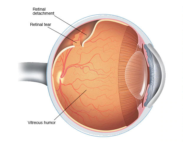
WHAT IS RETINAL DETACHMENT?
The separation of the retinal layer from the pigment epithelium layer to which it is anatomically attached due to a crack or hole is called rhegmatogenous retinal detachment.
How does retinal detachment occur?
There is a gel structure inside the eye that resembles egg white, which we call vitreous. The structure of the vitreous deteriorates with age. We perceive the deterioration in this gel structure as flies or strands of hair flying in front of our eyes. Sometimes the vitreous begins to separate from the retinal area it is attached to. During this time, symptoms such as flashes of light, soot, and flying flies may be observed in the eye. This situation may sometimes cause a piece of the retina to detach. This is when the retinal tear occurs. The situation can quickly turn into separation (detachment) in the retinal layers.
What are the symptoms of retinal detachment?
Some points in the visual field cannot be seen or vision may decrease. However, in retinal detachments where the visual center is not involved, the patient may not notice these defects because vision does not decrease.
What are the risk factors for retinal detachment?
The risk is higher in those with a family history of retinal detachment, high myopia, a history of eye trauma, and those who have had cataract surgery.
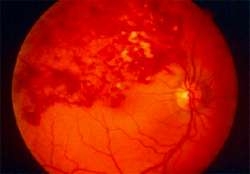
RETINAL VEIN OCCLUSION
What are the symptoms of the disease?
It mostly occurs with sudden vision loss at the age of 60-70. Depending on the location of the affected vein and the complications that occur, the decrease in vision may be mild or severe.
Are there types of retinal vein occlusion?
There may be occlusion in a single vein (branch of the retinal vein) or in the main vein (central retinal vein).
What are the risk factors for retinal vein occlusion?
Smoking, hypertension, diabetes, glaucoma (eye pressure) and various blood diseases that increase blood density are risk factors.
How is it examined?
Fundus is examined using fluorescein angiography (FFA) and OCT (Optical Coherence Tomography). These tests evaluate the amount and location of the obstruction, the presence of nutrition in the area, and whether there is fluid accumulation in the visual cortex.