HEALING

HEALING

During my long and challenging medical education, I used to think that my family wanted me to become a doctor and that I could turn back from this path at any time if I wished. As time passed and I climbed the steps of my training, I began to discover and understand myself. Initially, I kept my distance from the profession, but as I decided to embrace it, sharing my knowledge with my patients became increasingly enjoyable. Yes, healing was sacred, and I felt like I was observing the seedlings I planted in a pot take root in the soil—similarly, I believed my patients were healed through my hands. Every new medical student knows this: in the early years of being a doctor, you feel like the greatest physician. But as you progress, the ego that once roared like a tiger gradually shrinks, replaced by growing experience and confidence. This is exactly what happened to me…

I healed some of my patients with just a small touch. For others, I struggled greatly but couldn’t save them. This situation troubled me deeply. Why did nothing work for some of my patients? If I was the one providing the healing, why couldn’t I distribute it fairly? What was my role in all of this? Honestly, I pondered over this for a long time but couldn’t find an answer through mere thinking. Years passed, and as I discovered the philosophical side of the profession beyond its scientific aspect, I realized that I was not the one providing the healing. This realization was truly a shock to me.

Being a healer and a doctor requires being a conduit. If you are aware that you are not the source of the healing but merely a channel for it, you also benefit from that healing energy. That’s why, when a patient heals, the doctor heals too. Because when the healing flows through your conduit, a part of that goodness remains with you. This is why medicine is sacred and its teachings ancient.

When you are insulated, you cannot transmit either knowledge or healing. This is why great physicians know that knowledge also has its tax, and they always train students beneath them. In this way, knowledge and experience are passed on to the next generation like seeds. Transmission and sharing are the only stones on which your name will be written after you are gone.

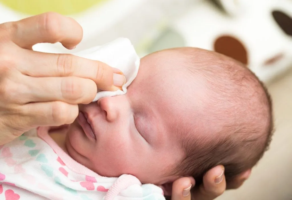
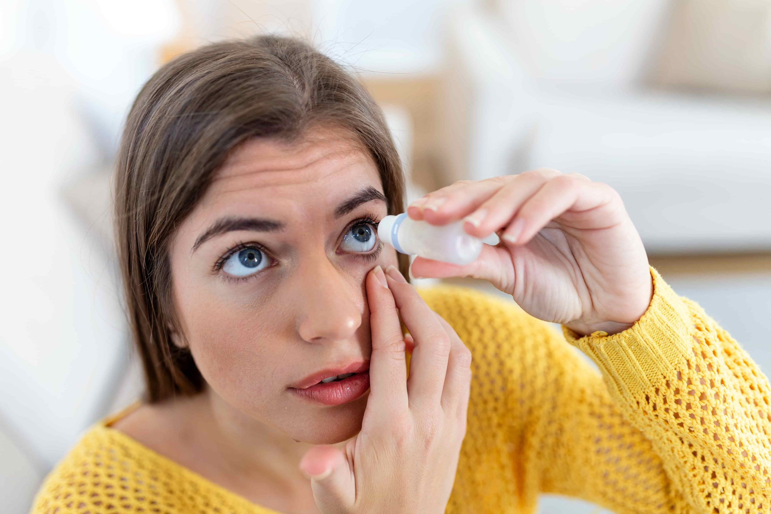
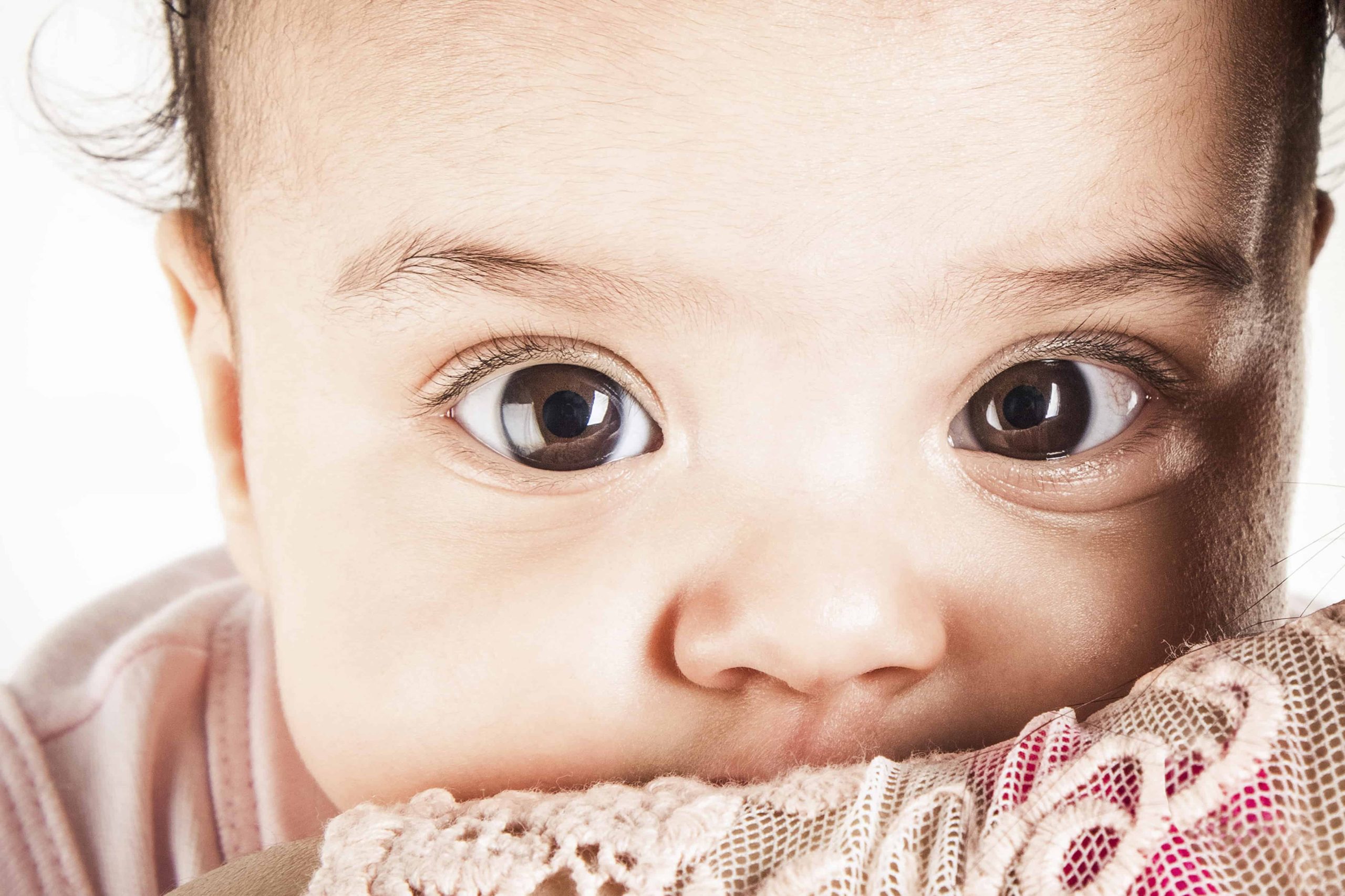
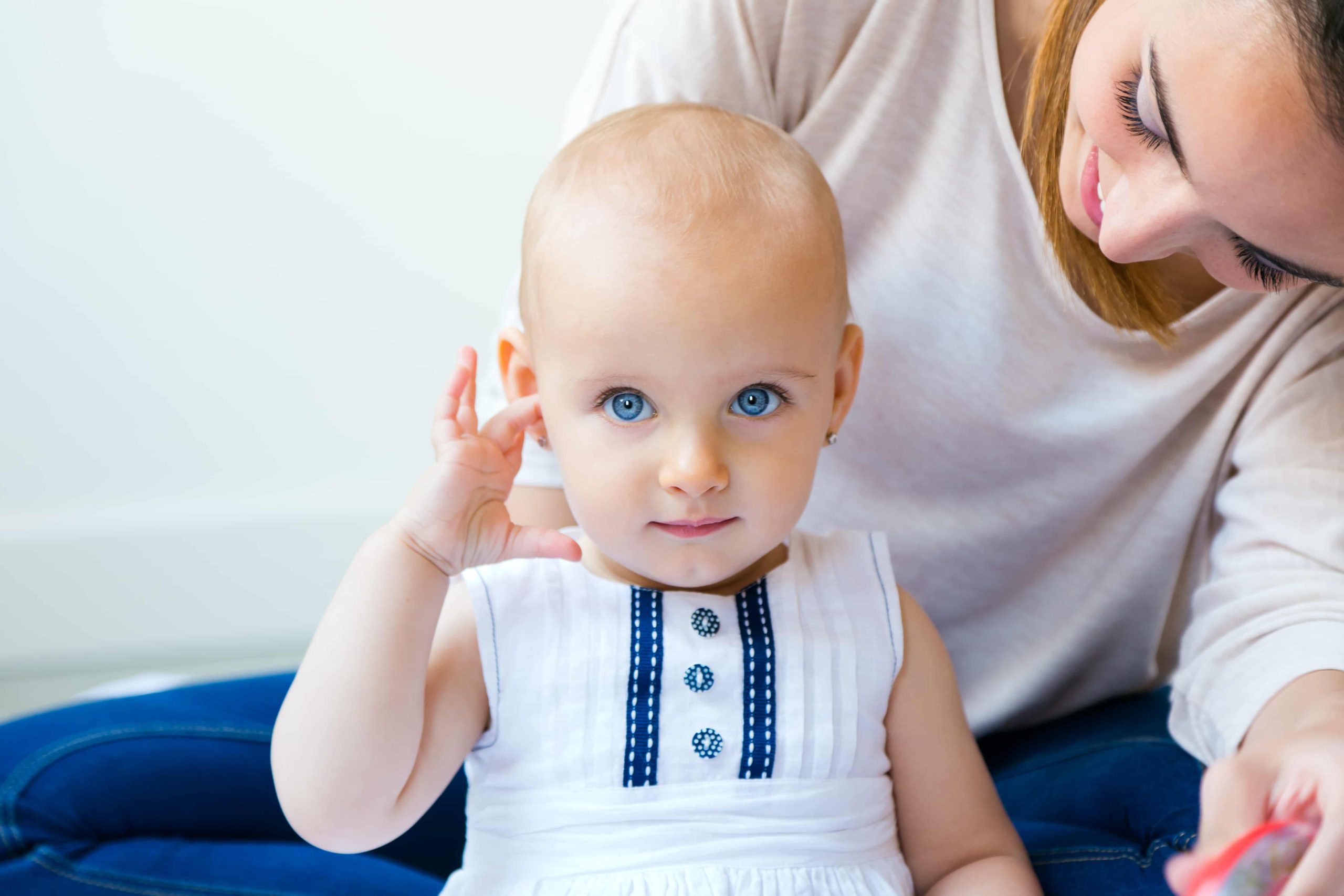
After I began to love my work, I always felt great joy as a woman practicing medicine. Because I believe that being a healer is inherently part of a woman’s nature. After all, every mother knows that when she kisses her child’s wound, she alleviates their pain and reduces their crying. Similarly, when you hug a friend tightly while they are upset, even if you haven’t found a solution to their problem, we all know through our feminine instincts that we are supporting them in their distress. Even a woman who has never heard the word empathy can easily understand the emotions of the person in front of her and respond effortlessly. That’s why being a female doctor sometimes simply means following your natural instincts.

Whatever work we engage in, perhaps when we, as women, lovingly accept our feminine side and begin to use the talents inherently given to us, we will step onto the main staircase that will elevate us.

Eye Watering and Discharge in Newborns

EYE WATERING AND DISCHARGE IN NEWBORNS

Eye watering in newborns is one of the most common issues we encounter in our outpatient practice. Therefore, it is important to educate families about this condition.

The underlying causes of eye watering in newborns can be identified through an ophthalmologist’s examination. I will try to explain the most common conditions causing eye watering, in order of their frequency.

The most common cause of intense discharge and crusting in newborns is the colonization of pathogens from the mother’s birth canal, leading to a severe eye infection. This condition is medically known as neonatal conjunctivitis. The discharge can be so thick that it sticks the eyelids together, and it often has a greenish-yellow appearance. It can occur in one or both eyes. The color and consistency of the discharge vary depending on the causative microorganism. The spectrum ranges from simple bacterial infections to more severe infections caused by Chlamydia or NeisseriaChlamydia and Neisseria infections produce a thicker, stickier discharge and respond more slowly to treatment. In such cases, collaboration with pediatricians is essential. Another important point is the evaluation of the mother’s birth canal. Since the infection is thought to be transmitted from the mother’s birth canal, it is crucial to ensure the mother receives appropriate treatment as well.

The second most common condition we see in the clinic is nasolacrimal duct obstruction (tear duct blockage). The nasolacrimal duct is a thin canal that carries tears from the eye to the nose. Congenital underdevelopment or the presence of a membrane within the duct can obstruct the natural flow of tears, causing them to accumulate and overflow. Initially, the tears are clear, but over time, the stagnant tears can become infected, leading to a more purulent discharge and crusting. This developmental issue is usually seen in one eye, though it can occasionally affect both eyes.

A much rarer cause of eye watering is congenital glaucoma. In this case, there is no discharge, but rather intense sensitivity to light and excessive tearing. Babies with congenital glaucoma cannot establish natural eye contact with their mothers due to this sensitivity. These infants typically have unusually large eye diameters and often have other congenital eye anomalies. Congenital glaucoma is an emergency condition in newborns, making accurate diagnosis crucial.

Macular Degeneration

Macular Degeneration

The medical name for macular degeneration is macular degeneration, which is a result of a circulatory problem that occurs due to the accumulation of metabolic residues in the retinal layer. For this reason, new vessels are formed in the macula, which is the central point of vision, and is the most important cause of vision loss over the age of 50.

Are there types of macular degeneration?
There are 2 types of the disease: wet and dry. The dry type progresses slowly. The wet type progresses more rapidly.

What are the symptoms of macular degeneration?
The symptoms of macular degeneration include broken or wavy vision, seeing the point you are looking at blurry and seeing your surroundings relatively better.

What are the risk factors for macular degeneration?
The most important ones are age and genetic predisposition. In addition to these, exposure to intense sunlight, smoking, obesity and hypertension are also effective.

How is macular degeneration diagnosed?
Diagnosis is made with eye angiography and eye tomography (OCT). If there is dye leakage outside the vessel or a new vessel is detected in angiography, the disease is classified as wet type. In OCT, detection of fluid within the retinal layers is in favor of wet type. Again, follow-ups are made with regular angiography and OCT.

Retinal Vein Occlusion

RETINAL VEIN OCCLUSION

What are the symptoms of the disease?
It usually occurs with sudden vision loss at the age of 60-70.
Depending on the location of the affected vein and the complications that occur, the decrease in vision may be mild or severe.

Are there types of retinal vein occlusion?
There may be occlusion in a single vein (branch of the retinal vein) or occlusion in the main vein (central retinal vein).

What are the risk factors for retinal vein occlusion?
Smoking, hypertension, diabetes, glaucoma (eye pressure) and various blood diseases that increase blood density are risk factors.

How is it examined?
It is examined using fundus fluorescein angiography (FFA) and OCT (Optical Coherence Tomography). These tests evaluate the amount and location of the blockage, the presence of nutrition in the area, and whether there is fluid accumulation in the visual center.

Retinal Detachment

WHAT IS RETINAL DETACHMENT?

The separation of the retinal layer from the pigment epithelium layer to which it is anatomically attached due to a crack or hole is called rhegmatogenous retinal detachment.

How does retinal detachment occur?
There is a gel structure inside the eye that resembles egg white, which we call vitreous.
The structure of the vitreous deteriorates with age. We perceive the deterioration in this gel structure as flies or hairs flying in front of our eyes. Sometimes the vitreous begins to separate from the retinal area it is attached to. During this time, symptoms such as flashes of light, soot, and flying flies can be observed in the eye. This situation can sometimes also cause a piece of the retina to detach. This is when a retinal tear occurs. The situation can quickly turn into separation (detachment) in the retinal layers.

What are the symptoms of retinal detachment?
Some points in the visual field cannot be seen or vision may decrease. However, in retinal detachments where the visual cortex is not affected, the patient may not notice these defects because vision is not reduced.

What are the risk factors?
The risk is higher in those with a family history of retinal detachment, those with high myopia, those with a history of eye trauma, and those who have had cataract surgery.

DRY EYE

DRY EYE

What is the use of tears?
Tears are of vital importance on the surface of the eye. Tears are necessary for the moistening, nourishment, purification of infections (both by washing away and with its antimicrobial effect), maintenance of corneal transparency and integrity, and maintenance of image quality on the retina.
What happens if tears are disrupted?
A decrease in tear secretion or a change in its components disrupts the balance on the surface of the eye. The deterioration in tear quality triggers inflammation on the surface of the eye, which results in damage to the layer called the epithelium that surrounds the surface of the eye in the form of a membrane. The opening of the nerve endings as a result of this epithelial damage causes discomfort in the eye, the need to blink frequently, and reflex tearing. While patients actually suffer from dry eyes, they may also apply to the clinic with excessive watering due to the paradoxical reflex tearing that develops. Here, we see that since the tears, which do not have sufficient content, cannot stay on the surface of the eye for the required time, the eye tries to eliminate this problem by producing more tears in order to keep these areas wet.

What triggers dry eyes?

Low humidity and excessive air flow increase tear loss through evaporation. Air-conditioned environments are effective in causing dry eyes for this reason. Long plane trips also cause dry eyes in a similar way. Computer use reduces the blink reflex and causes dry eyes. Caffeinated beverages can also cause dry eyes due to their diuretic (diuretic) effects. Therefore, it is useful not to strain the eyes, which are already exposed to an intensely air-conditioned environment, by taking caffeine during long plane trips. Air pollution and cigarette smoke are also effective in causing dry eyes.

Do medications trigger dry eyes?

Women, especially after menopause, have a higher potential to develop dry eyes due to the decrease in general body secretions. We know that dry eyes develop more frequently after eye surgeries such as LASIK and LASEK. We know that heavily used allergy medications, diuretics, and some antidepressant medications also cause dry eyes. Again, we know that eyelash root inflammation, or blepharitis, which we frequently see in clinical practice, also disrupts tear content and causes dry eyes.

Do contact lenses cause dry eyes?
It is known that contact lenses used for a long time also cause dry eyes.
Are there any diseases that accompany dry eyes?
A number of skin diseases, such as acne rosacea (rose rose) and rheumatic diseases (rheumatoid arthritis, lupus), can also cause dry eyes.

What can patients do to make their lives easier?
In the first step, the patient should be educated about dry eyes. Taking short breaks in computer use and blinking more often while using the computer are the simplest but most beneficial. Adjusting environmental factors is also important.
It is important to reduce the frequency of being in air-conditioned environments and, if possible, not to be in the opposite direction of the air-conditioning. Increasing water consumption is very important.

Staying away from caffeinated beverages, smoking and smoking environments helps to protect the tear film layer. A diet rich in greens and omega-rich foods is beneficial. It may be comforting for contact lens users to reduce the duration of use.

In patients with eyelash root inflammation, hot compresses and eyelash root cleaning with eyelash shampoo are meaningful for both the treatment of blepharitis and the treatment of secondary dryness.

Pupil whiteness

WHY DOES THE EYE OF A BABY HAVE WHITENESS?

We need a transparent layer inside the eye for vision. Light comes inward from the transparent glass-like layer at the front, which we call the cornea, and then it is directed into the structure we call the anterior chamber. It is filtered through the transparent lens tissue and reaches the vitreous tissue, which is like egg white and also transparent, that fills the eye. These areas are responsible for carrying photons to the retinal tissue, which perceives light.
A photon of light reaching the posterior segment is perceived by the cells in the retina and reaches the brain as data through the optic nerve. Loss of transparency in any of these ways disrupts the reflection of light inside the eye and creates a whitish reflex in the pupillary reflex instead of the traditionally expected reddish image, and this condition is called “leukocoria” in medical terms. It causes vision loss in the baby because it prevents light from reaching the eye. Therefore, diagnosis is very important. To briefly explain these; When there is a white pupil reflex, the first thing that comes to mind is congenital cataract.

The baby’s lens, which should be transparent, has become cloudy as if it were an old person, and the image has become blurry. This condition can sometimes be unilateral, sometimes bilateral.

The family’s history of cataracts in previous babies or in babies of cousins-uncles-aunts is very important because this condition is mostly transmitted genetically.

Another problem that impairs vision is the abnormal vascular structures and connective tissue bands that develop inside the normally transparent structure of the vitreous tissue. This condition is called persistent hyperplastic primary vitreous shortly PHPV. This structure is usually unilateral and can impair eye development in some babies and cause the eye to remain small. Congenital cataract, congenital glaucoma and retinal detachment, which occurs when the retinal layers are separated, may accompany the condition due to the abnormal development of the intraocular structures. We do not often encounter a family history in PHPV patients. One of the most feared conditions, retinoblastoma, is a tumor that develops in the retinal tissue. Tumor cells that develop abnormally in the area of ​​the retina that should see light and transmit it to the brain, both impair vision and threaten life. It can be unilateral and, unfortunately, sometimes bilateral. The presence of retinoblastoma in the family and especially in siblings should definitely be questioned. Genetic counseling is recommended for such families. In the event of this diagnosis, the baby should be seen by both an ophthalmologist and a pediatrician, because retinoblastoma is a life-threatening tumor.

The infection caused by toxocariasis, a parasite transmitted from dogs, in the retina and vitreous tissue is called toxocariasis. Infection in the retina can cause the death of rod and cone cells, while infection in the vitreous can cause both loss of transparency and the formation of band structures, pulling and separating the structures inside the eye, which is what we call detachment. Late diagnosis can cause problems leading to permanent blindness. Diagnosis can be made with eye examination and special laboratory tests.

The biggest problem of premature and low birth weight babies, premature retinopathy (ROP), moves from the retinal tissue into the vitreous, detaches the retina from its place and forms bands in the vitreous. This condition can cause permanent vision loss. The detached retina reveals itself by giving a white image in the eye and causes leukocoria. The risk is especially higher for premature babies under 32 weeks and babies with a birth weight under 1500 grams.

Although rare, Coats disease is a disease that should be kept in mind. It is a retinal vascular disease. Yellowish fluid leaking from the vessels in small foci accumulates between or under the retinal tissues and causes separation in the retinal tissue.
We know that this disease, which is mostly seen unilaterally, is more common in boys.
There are also rarer diseases seen in addition to these, but we have briefly explained the ones we encounter more frequently in practice. It is very important for an eye doctor to see a healthy red reflex in a baby due to the diseases we have listed above, so we can say that babies’ eye checks start from the moment they are born. I wish everyone healthy days.

Laziness of vision

WHAT IS LAZY VISUAL (AMBLYOPIA)?

LAZY VISUAL (AMBLYOPIA) is a disorder that occurs as a result of the visual center in the brain not developing due to insufficient visual stimuli reaching it, starting from an early age. If we summarize the causes of lazy vision under a few main headings;

1. Lazy vision due to refractive error is the most common group. Having a high number in one eye causes blurring of vision in that eye. Visual stimuli coming from two different eyes cause confusion in the visual center and the brain prefers to perceive the clearer vision. This is not actually a conscious choice, it is just a way the brain uses to survive. However, the fact that stimuli coming from the same eye are constantly used within a developing and growing system causes the visual system to be stimulated one-sidedly. As the visual center in the brain is stimulated, the networks between the neurons that provide vision will be triggered and vision will gradually begin to increase. Therefore, the visual center on the side that the eye with refractive error and blurred vision cannot stimulate lags behind in development. This means that the vision on the stimulated side gradually increases over time, but the vision on the unstimulated side does not develop.

2. Another reason is that the eye remains closed from the time the baby is first born. This can be thought of as not being able to get enough sunlight through a closed window. The closed condition is most frequently seen in cases of congenital eyelid droop, which we call congenital ptosis. Less frequently, the same problem can develop in congenital or later-developed eyelid masses located on the eyelid. Since the eyelid is droopy and the baby cannot lift it even if it wants to, that eye does not receive enough visual stimuli, and since the visual center in the brain cannot be stimulated sufficiently, amblyopia may develop due to the reasons we have explained above. Therefore, eyelid droop should not be perceived as just an aesthetic problem; it is useful to check whether refractive error and amblyopia have developed in that eye after the eyelid operation.

3. Strabismus is another cause of amblyopia. Children with strabismus usually cannot use their strabismus eye adequately, and preferentially use the eye that does not strabismus to see. Since the neuronal networks in the visual center stimulated by the strabismus eye do not develop sufficiently, vision in that eye will be limited. In strabismus, eye numbers (refractive errors), if any, are also corrected and the eyes help to send correct and sufficient vision signals to the brain.

4. Other less common causes of amblyopia include congenital cataracts, scars on the cornea that prevent vision, and opacities on the visual axis. In cases where the transparency on the visual axis is impaired, amblyopia may develop again because sufficient visual stimuli do not reach the visual cortex.

After amblyopia is diagnosed, it is very important for your child to be examined as often as your doctor recommends. Amblyopia is a disease that can be treated more easily with the family’s patience and support for the child.

Glocom

GLAUCOMA

Glaucoma is a disease that progresses with increased intraocular pressure. The optic nerve is damaged due to increased intraocular pressure. Since it is a slowly progressing disease, the classic symptom of a tunnel-shaped vision defect only occurs in the late stages. Since central vision is typically preserved until the advanced stages of the disease, it is unfortunately noticed by the patient very late.

Who is affected by glaucoma?
The risk of glaucoma increases in people over the age of 40.
Glaucoma may be related to genetics. The risk of development is higher in people with a family history of glaucoma. It is known that glaucoma is more common in those with migraine.
Glaucoma can develop in retinal detachment, eye tumors and eye infections such as chronic uveitis or iritis.
The frequency of glaucoma is approximately doubled in high myopes.
The risk of developing glaucoma is higher in those with diabetes.
Serious eye injuries can cause increased intraocular pressure.
Long-term cortisone use can cause glaucoma.
It is important for people with these characteristics to have regular eye examinations for early detection of optic nerve damage.

Does glaucoma occur in babies?

Yes, the type called congenital glaucoma is seen in the newborn period and can cause a large eye appearance called buphthalmus due to excessive expansion in the elastic eye structure.

While drug treatment is primarily administered to adults, the primary treatment in babies is surgery.

How does a glaucoma crisis occur?
It usually occurs in patients with a narrow angle in the eye, when the angle closes and blood pressure suddenly increases. The eye becomes red, painful and vision is blurred. Emergency drug treatment applied at this stage prevents permanent damage to the eye. After the emergency situation is corrected, the patient is given drops that he/she can use continuously and after the eye calms down, laser treatment can be applied according to the suitability of the doctor.
How is glaucoma monitored?
Although it varies depending on the condition of the eye and the patient, patients are usually evaluated by an ophthalmologist every 3-4 months. Glaucoma tests are performed and the patient is informed about the condition of the eye.

Could My Baby Have Eye Pressure?

CAN MY BABY HAVE EYE TENSION?
Congenital eye tension is a rare disease that can be seen in babies. Due to increased eye pressure, the very fresh intraocular tissues are stretched and cracks occur in the corneal tissue, which acts like glass in the front structure of the eye. With the addition of edema, the corneal tissue loses its transparency. This situation both reduces vision and creates excessive watering and abnormal sensitivity to light.

A baby whose cornea is edematous and whose intraocular pressure increases squeezes his eyelids excessively and has great difficulty opening his eyes. In babies whose condition is not recognized and whose diagnosis is delayed, cracks in the transparent corneal tissue become fixed and create a blurry image. Since the eye with eye tension will see blurry, its ability to follow decreases and it cannot fixate on a point and focus. If we think of the eye as a balloon, the structure of the eye gradually grows as the intraocular pressure increases over time. The eye becomes much larger than it should be. This condition is especially understood more quickly if there is congenital glaucoma in one eye, as asymmetry will develop.

In the presence of bilateral glaucoma, it is necessary to be much more careful since both eyes will grow abnormally. This excessive growth in the eye may regress after the intraocular pressure returns to normal, but eyes that have remained in this state for a very long time may still remain somewhat large. Increased eye diameter may cause changes in the number in the future.

In advanced cases where glaucoma is not noticed, the eye diameter gradually increases and the eye structure begins to deteriorate. The pressure on the optic nerve can cause degeneration and cause permanent deterioration in the optic nerve. Congenital glaucoma is often accompanied by congenital anomalies of other structures in the eye. This condition is understood during the examination by the ophthalmologist when you take your baby for a routine check-up.

1 2

Search

+